Researchers adapted NMR protocols to outline chemical structures in cell wall material that is a potential source of valuable compounds.
The Science
Nuclear magnetic resonance (NMR) is a tool scientists can use to identify chemical compounds, including those locked up in complex plant tissue. The NMR machine produces strong magnetic fields and uses radio waves to cause certain atoms to give off signals the machine can detect. The frequencies of these signals vary based on the chemical environment and how close they are to other atoms. By analyzing patterns in the signals, researchers can piece together a molecule’s structure. Scientists use NMR spectroscopy to study lignin, a part of the plant cell wall that provides structure. Lignin is a polymer, a matrix of smaller ring-shaped molecules that are potentially valuable. These aromatic units are linked by various chemical bonds in ways that vary by plant species and even tissue type. Researchers need to understand the structure and chemical bonds in order to break lignin apart in ways that yield usable compounds.
Nearly 99% of the carbon on Earth is carbon-12, which has an even number of protons and neutrons and doesn’t show up on NMR scans. A little more than 1% is carbon-13 (13C), a variant with one extra neutron that does show up on NMR scans. Enriching biomass to 10% to 15% 13C sharpens the picture and makes it easier to identify small structures that would otherwise be invisible. Some researchers have grown fully-labeled 13C plants, which they assumed would yield even higher sensitivity. But when every carbon atom is labeled they interact, causing the signals to split or fade, blurring the picture.
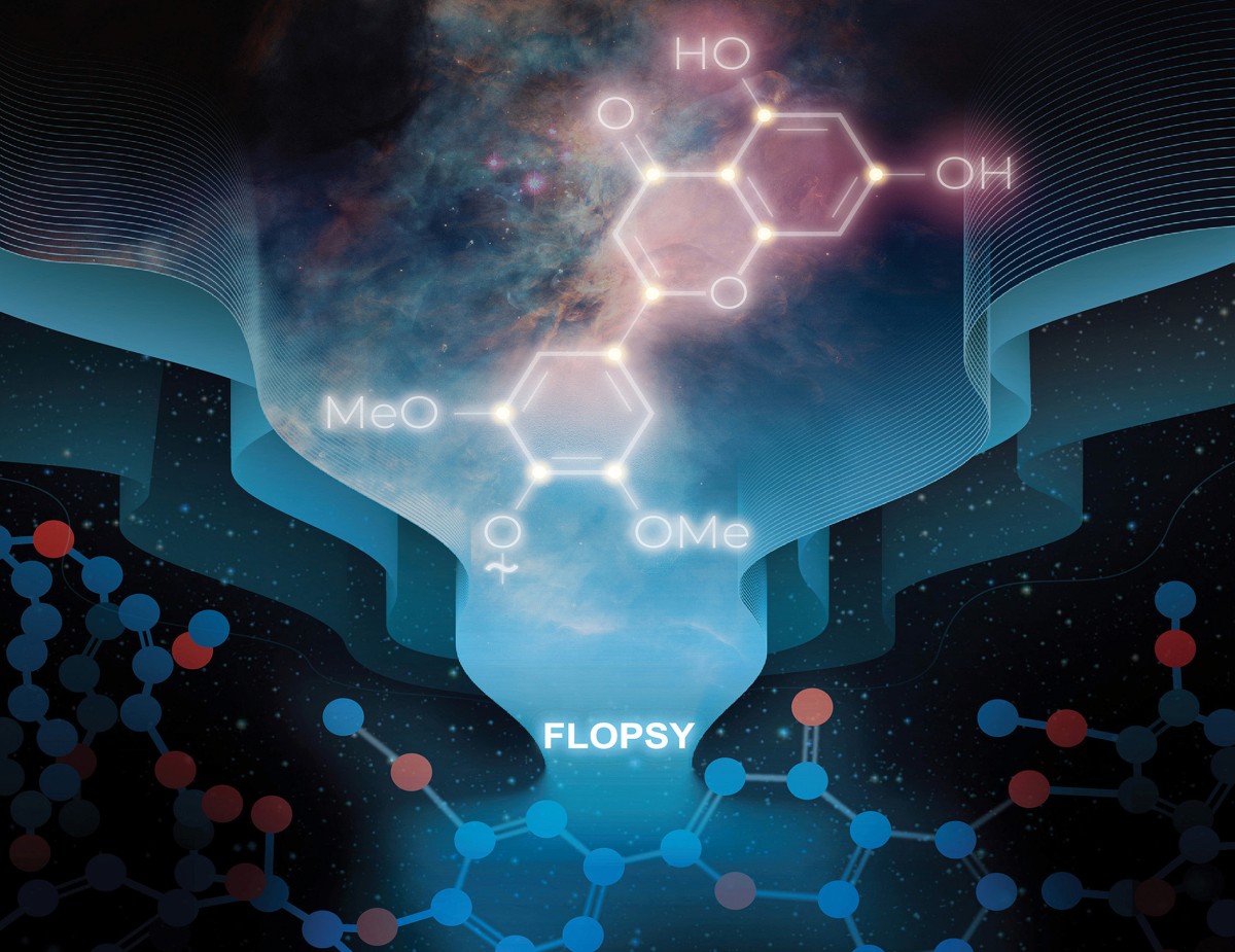
A team of international researchers led by the Great Lakes Bioenergy Research Center used alternate methods to get around the problem with fully-labeled biomass. One experiment used a hybrid approach known as CT-HSQC-TOCSY to provide a clearer picture of carbon–hydrogen pairs and how they are connected. A technique known as FLOPSY takes advantage of the carbon–carbon interactions to produce a sort of skeleton map of carbon atoms, even when they are part of a complex polymer like lignin. It also reveals small “pendent” molecules that would be difficult to identify with other techniques, enabling researchers to verify the presence of one such compound for the first time.
The Impact
Non-food plant material, such as crop residue or purpose-grown energy crops, can be a source of domestically grown feedstocks for fuels and chemicals used to make plastics, medicines, and other essential products. However, complex chemical structures make it challenging to break plant biomass into useful parts. This study demonstrates how two NMR experiments can be applied to deliver clearer and more detailed pictures of complex plant polymers, which can help researchers better understand what compounds are in plant biomass as well as how they are arranged.
Summary
NMR spectroscopy is the premier method for profiling the composition and structure of the plant cell wall polymer lignin. Informative solution-state spectra can be generated from polysaccharide and lignin components even in finely milled cell-wall materials. Cultivating plants in 13C-enriched carbon dioxide to a level of 10-15% has proven advantageous for enhancing sensitivity (signal to noise) of spectra and facilitating rigorous authentication of minor components. Researchers have produced near-100% 13C-enriched biomass. Counterintuitively, standard NMR analyses of such labeled cell wall material did not yield higher sensitivity due to complications arising from 13C–13C coupling.
This research showed that constant-time HSQC (CT-HSQC) experiments mitigate these issues, delivering enhanced sensitivity using 13C-enriched poplar, sorghum, and maize straw. A rarely-reported CT-HSQC-TOCSY experiment allows the proton coupling network to deliver much of the same value as the parent experiment on unlabeled or 10-15% 13C-enriched biomass polymers, but with enhanced sensitivity. A C–C FLOPSY experiment takes advantage of fully 13C-labeled materials for mapping extensive carbon networks in complex biomass polymers. This first application of the methods revealed various pendent groups (tricin units, cis- and trans-p-coumarates, and p-hydroxybenzoates) that adorn lignins, and the cis- and trans-ferulates on arabinoxylan polysaccharides are exquisitely revealed in spectra from isolated lignins or whole-cell-wall materials. Spectra of fully-13C-labeled sorghum leaf cell wall samples enable the previously unassigned cis-ferulate (cF) peaks to be elucidated, validating recent speculation regarding their likely presence.
The C–C FLOPSY experiment has significant additional value for studies on fully 13C-labeled biomass due to its superior applicability to readily prepared cell wall samples. Its shorter pulseprogram profile enables the acquisition of impressive spectra from rapidly relaxing samples for which experiments useful for unlabeled materials simply fail.